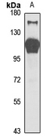
Western blot analysis of Striatin-3 expression in mouse brain (A) whole cell lysates. (Predicted band size: 77; 87 kD; Observed band size: 110 kD)
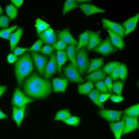
Immunofluorescent analysis of Striatin-3 staining in MCF7 cells. Formalin-fixed cells were permeabilized with 0.1% Triton X-100 in TBS for 5-10 minutes and blocked with 3% BSA-PBS for 30 minutes at room temperature. Cells were probed with the primary antibody in 3% BSA-PBS and incubated overnight at 4 °C in a hidified chamber. Cells were washed with PBST and incubated with a Alexa Fluor 488-conjugated secondary antibody (green) in PBS at room temperature in the dark.