KLH-conjugated synthetic peptide encompassing a sequence within the N-term region of human BMX. The exact sequence is proprietary.
Description:
Rabbit polyclonal antibody to BMX
Uniprot:
P51813,P97504
BiowMW:
Refer to Figures
Buffer:
Liquid in 0.42% Potassium phosphate, 0.87% Sodium chloride, pH 7.3, 30% glycerol, and 0.01% sodium azide.
Storage:
Store at 4°C short term and -20°C long term. Avoid freeze-thaw cycles.
Note:
For research use only, not for use in diagnostic procedure.
Alternative Name:
BMX; Cytoplasmic tyrosine-protein kinase BMX; Bone marrow tyrosine kinase gene in chromosome X protein; Epithelial and endothelial tyrosine kinase; ETK; NTK38
Data:
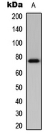
Western blot analysis of BMX expression in mouse brain (A) whole cell lysates.
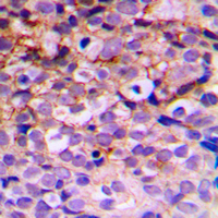
Immunohistochemical analysis of BMX staining in human breast cancer formalin fixed paraffin embedded tissue section. The section was pre-treated using heat mediated antigen retrieval with sodium citrate buffer (pH 6.0). The section was then incubated with the antibody at room temperature and detected using an HRP conjugated compact polymer system. DAB was used as the chromogen. The section was then counterstained with haematoxylin and mounted with DPX.
Immunofluorescent analysis of BMX staining in A549 cells. Formalin-fixed cells were permeabilized with 0.1% Triton X-100 in TBS for 5-10 minutes and blocked with 3% BSA-PBS for 30 minutes at room temperature. Cells were probed with the primary antibody in 3% BSA-PBS and incubated overnight at 4 °C in a hidified chamber. Cells were washed with PBST and incubated with a DyLight 594-conjugated secondary antibody (red) in PBS at room temperature in the dark. DAPI was used to stain the cell nuclei (blue).